Pifithrin-μ 是p53和HSP70的抑制剂,具有抗肿瘤和神经保护作用。
产品描述
Pifithrin-μ is an inhibitor of p53 and HSP70, with neuroprotective and antitumor activity.
体外活性
Pifithrin-μ interferes with p53 binding to mitochondria and inhibits rapid p53-dependent apoptosis of primary cell cultures of mouse thymocytes in response to gamma radiation. [1] Pifithrin-μ, as an inhibitor of inducible HSP70, significantly inhibited cell viability with IC50 values ranging from 2.5 to 12.7?μM in acute myeloid leukemia (AML) and acute lymphoblastic leukemia (ALL) cell lines, as well as in primary AML blasts. Pifithrin-μ is also highly active in primary AML blasts with a median IC50 of 8.9?μ M(range 5.7-37.2 ?μ M) and induces apoptosis and cell cycle arrest in a dose-dependent fashion. In addition, Pifithrin-μ also increases the active form of caspase-3 and reduces AKT and ERK1/2 in NALM-6 cells. [2] Pifithrin-μ increases Annexin V(+) cells in both caspase-dependent and -independent manners and promotes TRAIL-induced apoptosis and arrested cancer cell growth. [3]
体内活性
Pifithrin-μ (40 mg/kg, ip) produces protective effect from p53-dependent apoptosis in C57B1/6J mice of 8 or 9 Gy of total body gamma radiation. [1] In a xenograft mouse model, Pifithrin-μ significantly enhances the inhibition of TRAIL on MiaPaca-2 tumor growth. [3]
激酶实验
In vitro Kinase Assays: Kinase assays for CDK1, CDK2 and GSK3-β are all carried out in a radiometric filter binding format. Assays for CDK5 are in DELFIA format and for CDKs 4 and 6 in ELISA format. For CDKs 1 and 2, the relevant CDK and 0.12 μg/mL Histone H1 are incubated in 20 mM MOPS, pH 7.2, 25 mM β-glycerophosphate, 5 mM EDTA, 15 mM MgCl2, 1 mM sodium orthovanadate, 1 mM DTT, 0.1 mg/mL BSA, 45 μM ATP (0.78 Ci/mmol) and different concentrations of AT7519 for 2 or 4 hours respectively. For GSK3-β, the relevant enzyme and 5 μM glycogen synthase peptide 2 along with 10 mM MOPS pH 7.0, 0.1 mg/mL BSA, 0.001% Brij-35, 0.5% glycerol, 0.2 mM EDTA, 10 mM MgCl2, 0.01% β-mercaptoethanol, 15 μM ATP (2.31 Ci/mmol) and different concentrations of AT7519 are incubated for 3 hours. Assay reactions are stopped by adding an excess of orthophosphoric acid and filtered using Millipore MAPH filter plates. The plates are then washed, scintillant added and radioactivity measured by scintillation counting on a Packard TopCount. For CDK5, CDK5/p35 and 1 μM of a biotinylated Histone H1 peptide (Biotin-PKTPKKAKKL) are incubated in 25 mM Tris-HCl, pH 7.5, 2.5 mM MgCl2, 0.025% Brij-35, 0.1 mg/mL BSA, 1 mM DTT, 15 μM ATP and different concentrations of AT7519 for 30 minutes. Assay reactions are stopped using EDTA, transferred to Neutravidin-coated plates and phosphorylated peptide quantified by means of a rabbit phospho-cdk1 substrate polyclonal antibody and DELFIA europium-labelled anti-rabbit IgG secondary antibody using time-resolved fluorescence at λex=335 nM, λem=620 nM. For CDK 4 and 6 assays, plates are coated with GST- pRb769-921 and blocked with Superblock. CDK4 or 6 is incubated with 15 mM MgCl2, 50 mM HEPES, pH 7.4, 1 mM DTT, 1 mM EGTA, pH 8.0, 0.02% Triton X-100, 2.5% DMSO and different concentrations of AT7519; the reaction is initiated by addition of ATP. After 30 minutes, reactions are stopped by the addition of 0.5 M EDTA pH 8.0.Plates are then washed and incubated for one hour with the primary antibody (anti- p-Rb Serine 780) diluted in Superblock followed by secondary antibody (alkaline phosphatase linked anti-rabbit) for a further hour. Plates are developed using the Attophos system and fluorescence read on a Spectramax Gemini plate reader at excitation 450 nm and emission 580 nm. In all cases, IC50 values are calculated from replicate curves, using GraphPad Prism software.
细胞实验
The number of attached cells is estimated by staining with 0.5% methylene blue and measuring optical density using a Multiscan Ascent microplate reader. Cell viability is determined in suspension of short-term culture of primary thymocytes by staining them with 0.1% trypan blue2 or by analysis of annexin- or propidium iodide (PI)-positive cells by FACScan. (Only for Reference)
Cas No.
64984-31-2
分子式
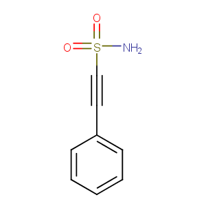
C8H7NO2S
分子量
181.21
别名
NSC 303580;PFTμ;2-Phenylethynesulfonamide
储存和溶解度
Ethanol:13.1 mg/mL (100 mM)
DMSO:13.1 mg/mL (100 mM)
Powder: -20°C for 3 years
In solvent: -80°C for 2 years
 m.cnreagent.com